Scan Findings: Terms Explained


AORTA

AORTIC VALVE

LEFT ATRIUM

PULMONARY ARTERIES

RIGHT VENTRICLE

RIGHT ATRIUM

LEFT VENTRICLE

MITRAL VALVE


TRICUSPID VALVE


PULMONARY VALVE
What do your results mean?
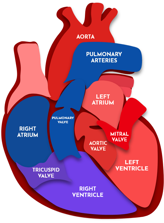
This interactive diagram of the heart explains the anatomy assessed during your scan.
The heart is a muscular pump, with two filling chambers (atria) and two pumping chambers (ventricles) all separated by four valves. Blood enters the right side of the heart (the right atrium) via the body’s large veins – the superior and inferior vena cava. The blood descends through the tricuspid valve into the right sided pumping chamber (the right ventricle), where it passes through the pulmonary valve into the lungs to collect oxygen. The blood returns from the lungs into the left atrium, where it descends through the mitral valve into the left ventricle. The left ventricle is the most powerful chamber, and is responsible for pumping blood through the aortic valve into the body’s largest blood vessel – the aorta – and off around the body to where it is needed.
Most cardiac abnormalities are classified according to the following (increasing) grades of severity:
Trace or Trivial
Trace or Trivial abnormalities are of no concern; these subtle changes can be seen in otherwise perfectly normal hearts.
Mild
Mild abnormalities are often in keeping with the normal ‘wear and tear’ of the heart, and only occasionally need specialist follow-up.
Moderate
Moderate abnormalities – especially pertaining to valves – are likely to need regular monitoring to check for progression over time.
Severe
Severe abnormalities usually warrant specialist referral. In the case of the heart valves, severe abnormalities may necessitate valve replacement surgery.


Even normal scan reports may contain unfamiliar terminology: find out what it all means below
This list includes common medical terms and phrases used to transform your scan images into a written report.
Acoustic windows
The ease with which an ultrasound probe can collect images of the heart. For example, patients with chest wall or spinal conditions may have ‘limited’ or ‘challenging’ acoustic windows, and as such certain areas of the heart may not be fully visualised with ultrasound.
Akinesis
No movement.
Anterior
At the front.
Bicuspid
A valve made up of two segments (this term usually refers to a genetically abnormal aortic valve, which should have three segments).
Calcification
Calcium which has built up on the valves over time (like limescale in a washing machine).
Diastolic function
An assessment of how efficiently the heart fills; diastolic dysfunction is suggestive of a stiff heart (this can be normal in older people).
Dilated
Enlarged (usually describing a chamber or blood vessel).
Dyskinesis
Abnormal, asynchronous movement.
Eccentric
Usually refers to regurgitation. Eccentric regurgitation suggests a leak through a valve at an abnormal angle, whereas concentric regurgitation suggests a leak straight through the middle of the valve.
Effusion
A collection of fluid around the heart or the lung.
Ejection fraction
A percentage estimate of the pumping function of the heart. At rest, around 55% is a normal ejection fraction. (see above)
Hyperdynamic
This normally refers to systolic function, and describes a heart that is pumping more forcefully than expected.
Hypertrophy
Thickened (usually describing the heart muscle). This can be further described as 'concentric', in which all the heart muscle is thickened equally, or 'asymmetric', in which certain areas of the heart are thicker than others.
Lateral
Towards the side.
Leaflet
Leaflets (or 'cups') are the individual segments that make up the heart valves.
Mass
A lump or collection of abnormal material.
Medial
Towards the middle.
Patent foramen ovale
We are all born with a small hole in the heart which connects both atria. In some people, this hole fails to close, which can cause a shunt of blood. (see below)
Posterior
At the back.
Preserved
Not significantly reduced (i.e essentially normal).
Prolapse
A valve that doesn't close properly.
Regional wall motion abnormality
An area (or 'segment') of the heart muscle with reduced movement.
Regurgitation
A leak back through a valve after it closes.
Rheumatic
A characteristic stiffening of a valve suggestive of previous rheumatic fever.
Sclerotic
Minor calcification on a valve's leaflets.
Shunt
An abnormal flow of blood from one side of the heart to the other, which usually occurs through a hole in the middle of the heart.
Stenosis
Abnormally narrow valve causing some obstruction of blood flow.
Systolic function
The pumping function of the heart's ventricles.
Thrombus
A blood clot.

SPECIFIC ABNORMALITIES
Read detailed descriptions of common cardiac abnormalities seen on ultrasound.
Read MoreBook Your Heart Scan Today